
Amritsar Eye Clinic is well equipped to deal with various forms of Uveitis. Apart from basic facilities for the diagnosis of Uveitis like high magnification slit-lamps and indirect ophthalmoscopy, the centre offers the following to its uvea patients.
Fundus Photography
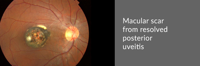
For visualization of the posterior part of the eye, often termed the Fundus.
Fundus Fluorescein Angiography (FFA)
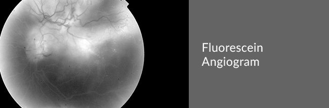
Helps to study the Fundus in greater detail, with particular attention to the vasculature. Fundus photographs are taken after intravenous injection of a special dye.
Optical Coherence Tomography (OCT)
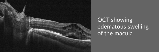
For the detailed study, at the microscopic level, of the macula, optic disc and the retinal nerve fibre layer (RNFL). It is of particular importance in the detection of Macular Oedema.
Ultrasound B-scan
Gives a 2-D or cross-sectional image of the entire eye-ball. It helps in detecting complications like intra-ocular tumours, Retinal Detachment, swellings of the choroid and retina, etc.
Complications of Uveitis like secondary cataract and secondary glaucoma can also be well managed.